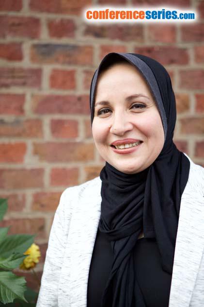
Naglaa Kamal Idriss
Assiut University, Egypt
Title: Comparison of treatment proficiency of intravenous and intrasplenic injection of the bone marrow-derived mesenchymal stem cell in CCL4-induced white albino rat liver fibrosis
Biography
Biography: Naglaa Kamal Idriss
Abstract
Background: Human umbilical cord blood (UCB) cells and rat bone marrow mesenchymal stem cells (BM-MSCs) have many advantages as grafts for cell transplantation.
Aim: The aim of this study was to evaluate the treatment effects of rat bone marrow-derived mesenchymal stem cells (BM-MSCs) on rat liver fibrosis induced by carbon tetrachloride. Intrasplenic and intravenous transplantations were examined to evaluate the effects of different injection routes on the liver fibrosis model at 12 weeks after transplantation.
Methods & Results: Experimental animals include 24 male white albino rats were 4 weeks old, weighing between 130 and 150 g. Liver fibrosis was induced by subcutaneous injection of carbon tetrachloride (CCl4) at a dose of 0.2 ml/100 g body weight of 40 ml/L CCl4 dissolved in equal volume of castor oil. The injection was given twice weekly for 6 week rats were divided into the following groups: G1 (Control group): 6 rats received 0.2 ml/100 g body weight of castor oil twice weekly for 6 weeks. G2 (CCl4 group): 6 rats received 0.2 ml/100 g body weight of CCl4. Liver fibrosis was determined by histopathological examination. G3 (CCl4/BM-MSCs group): 6 rats received CCl4 as previous. The rats were infused with 107 BM-MSCs/rat intravenously (through tail vain) and scarified after 3 months. G4 (CCl4/BM-MSCs group): 10 rats received CCl4 as previous and followed by injection of 107 BM-MSCs intrasplenic and scarified after 3 months. At 4, 8 and 12 weeks from stopping CCl4 and administration of stem cells, venous blood was collected from the retro-orbital vein to assess serum albumin and alanine transaminase (ALT). All rats were sacrificed with CO2narcosis and the liver tissue was harvested for histopathological examination and real time PCR analysis. Isolation and Culture of BM-MSCs. Cultured MSCs were confirmed by morphology, labeling Stem Cells with GFP. Serum ALT and albumin were assessed using colorimeter kits according to manufacture instructions.Histopathological examination liver tissues were collected and divided into two sections. The first section was assessed for tracing of injected labeled cells with GFP. The second section was washed with PBS and fixed overnight in 40 g/L paraformaldehyde at 4 °C for evaluation of fibrosis. Real Time PCR (qRT-PCR) for Quantitative Expression of IL-6, IL1-β, CK18, INF-γ and HGF. Western blotting for human SIRT-1: The results of the blots are presented as direct comparisons of the area of the apparent bands in autoradiographs and quantified by densitometry using the Bio- Rad Image software. ELISA: Connective tissue growth factor(pg/ml) was assessed.
Conclusion: Notably, there were no differences in treatment effects between intravenous and intrasplenic administrations. The IV injection group had significantly different (p<0.05) serum connective tissue growth factor levels compared with the intrasplenic injection group. However, liver serum markers and liver histology classification of both groups showed no differences (p>0.05). Considering safety, BM-MSC transfusion via a peripheral vein is a potential method for liver fibrosis treatment. In consideration of safety, we suggest transfusion of bone marrow-derived mesenchymal stem cells via a peripheral vein as a potential method for liver fibrosis treatment.
Speaker Presentations
Speaker PPTs Click Here


